
-
Mail us:
editor@raftpubs.org
Indexing & Abstracting
Full Text
Case BlogDOI Number : 10.36811/jcri.2021.110019Article Views : 34Article Downloads : 24
Unilateral pneumocephalus: A rare case
Abdulwahab Alahmari
Radiology Specialist, Radiology Department, Al-Namas General Hospital, Ministry of Health, Al-Namas City, Saudi Arabia
*Corresponding Author: Abdulwahab Alahmari, Radiology Specialist, Radiology Department, Al-Namas General Hospital, Ministry of Health, Tel: +966562428716; Email: afaa99@hotmail.co.uk
Article Information
Aritcle Type: Case Blog
Citation: Abdulwahab Alahmari. 2021. Unilateral pneumocephalus: A rare case. J Case Rept Img. 3: 01-09.
Copyright: This is an open-access article distributed under the terms of the Creative Commons Attribution License, which permits unrestricted use, distribution, and reproduction in any medium, provided the original author and source are credited. Copyright © 2021; Abdulwahab Alahmari
Publication history:
Received date: 26 December, 2020Accepted date: 15 January, 2021
Published date: 19 January, 2021
Abstract
The aim of this paper is to share a rare finding that is unreported in any peer reviewed journal before to the best of our knowledge. Pneumocephalus is characterized by the classic “Mount Fuji” sign. This case will present a patient suffers from a frontal and nasal bone fracture on one side that caused a unilateral pneumocephalus.
Keyword: Pneumocephalus; Mount Fuji Sign; Computed Tomography; Road Traffic Accident
Introduction
Pneumocephalus is a condition where air entered (in one way and can’t escape) the subdural space then it makes mass effect on the brain parenchyma. There is a posted case of unilateral tension pneumocephalus on radiology cases cite that has similar findings [1]. Otherwise, no published case with similar findings have been published in a peer reviewed journal.
Case Report
A twenty-one-year-old male patient came to emergency department after he had a road traffic accident (RTA), his nose was leaking cerebrospinal fluid (CSF), and the patient suffer from poly-trauma. A PAN CT scan was done for him. The CT showed a frontal and nasal bone fracture on the left side that caused unilateral pneumocephalus. The pneumocephalus did not show the classic “Mount Fuji” sign and the CT scan revealed some air bubbles in the subarachnoid space see (figure 1-29). The PAN CT and x-rays for the affected limbs were done which revealed multiple bilateral osteochondroma known as diaphyseal aplasia -which is a rare autosomal disorder-affecting the entire skeletal system (figure 30-36). The patient complains of frozen shoulders and can’t move his upper limbs. The patient is stable and has severe headache. A previous head CT scan showed that the patient before the accident have enlarged fourth ventricle, occipital fossa, basilar artery calcification (is not provided in this case report).

Figure 1: A frontal bone fracture extended to the nasal bone on the left side (bone window).
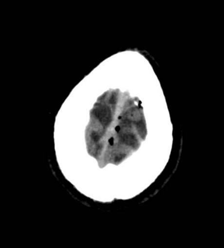
Figure 2: Air bubbles in the left subarachnoid space.
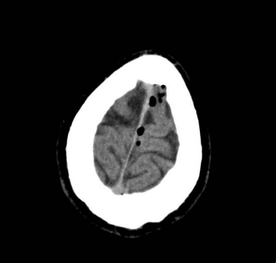
Figure 3: Air bubbles in the subarachnoid space on the left side.
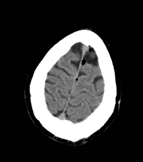
Figure 4: Air bubbles in the left side and near the midline.
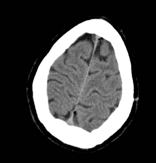
Figure 5: Air bubbles located in the front and the back of the superior part of the brain.
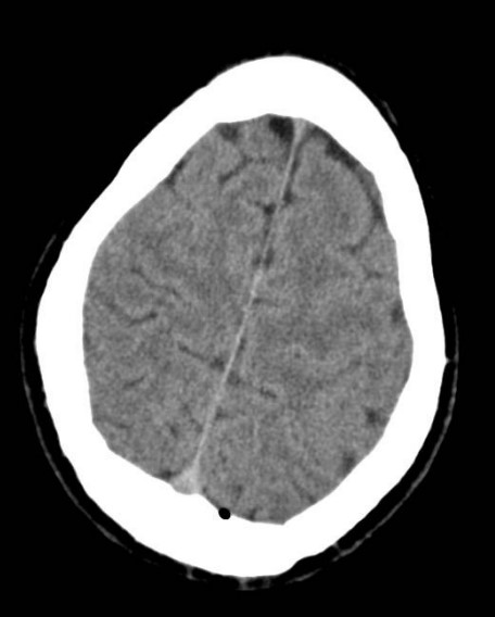
Figure 6: Air bubble in the posterior part of the brain (left side).
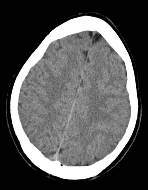
Figure 7: Air bubble located posterior to the left cerebral hemisphere.
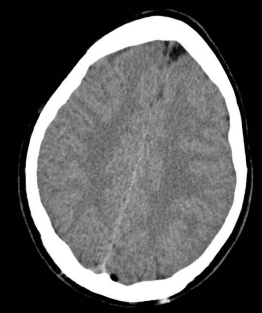
Figure 8: Air bubble posterior to the left cerebral hemisphere.
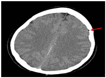
Figure 9: Air bubble located lateral to the left cerebral hemisphere.
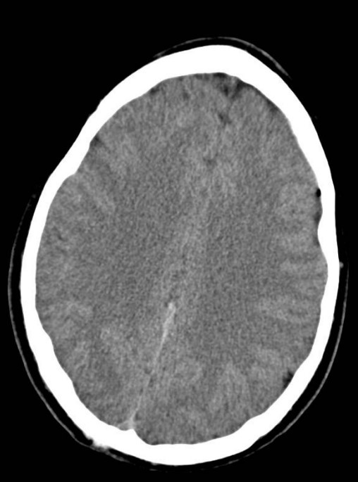
Figure 10: Air bubble located lateral to the left cerebral hemisphere.

Figure 11: Air accumulation anterior to the left cerebral hemisphere.

Figure 12: Air accumulation anterior to the left cerebral hemisphere.

Figure 13: Unilateral pneumocephalus (CT scan, axial section, brain window).
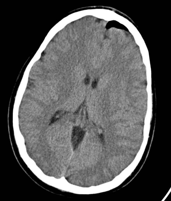
Figure 14: Unilateral pneumocephalus (CT scan, axial section, brain window).
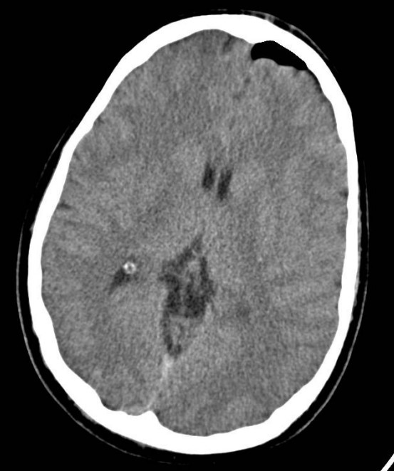
Figure 15: Unilateral pneumocephalus (CT scan, axial section, brain window).
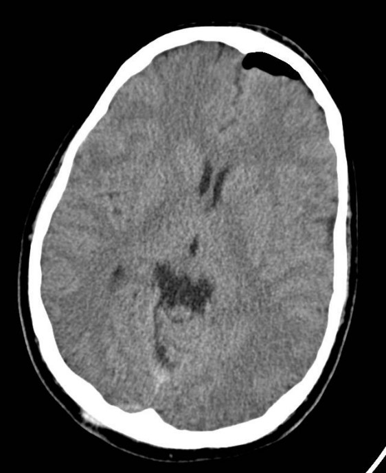
Figure 16: Unilateral pneumocephalus (CT scan, axial section, brain window).

Figure 17: Unilateral pneumocephalus (CT scan, axial section, brain window).
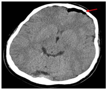
Figure 18: Unilateral pneumocephalus (CT scan, axial section, brain window).
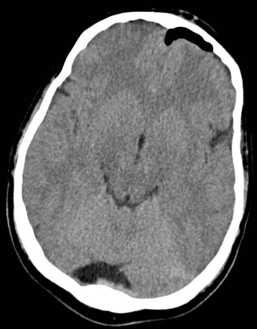
Figure 19: Unilateral pneumocephalus on the left side with no involvement from the right side (CT scan, axial section, brain window).
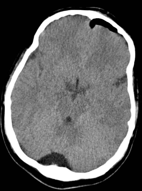
Figure 20: Unilateral pneumocephalus and enlarged occipital fossa (CT scan, axial section, brain window).
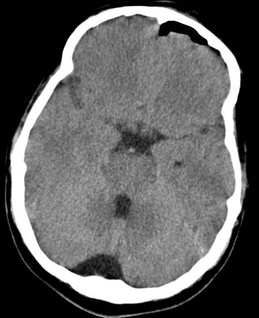
Figure 21: Unilateral pneumocephalus and enlarged occipital fossa and fourth ventricle (CT scan, axial section, brain window).
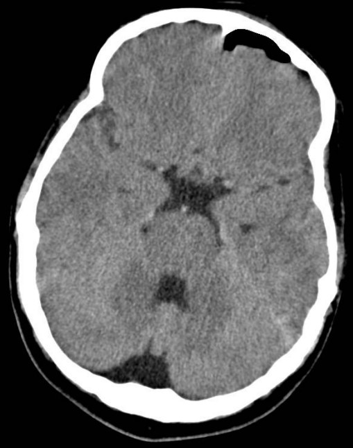
Figure 22: Unilateral pneumocephalus and enlarged occipital fossa, fourth ventricle, and suprasellar cistern (CT scan, axial section, brain window).
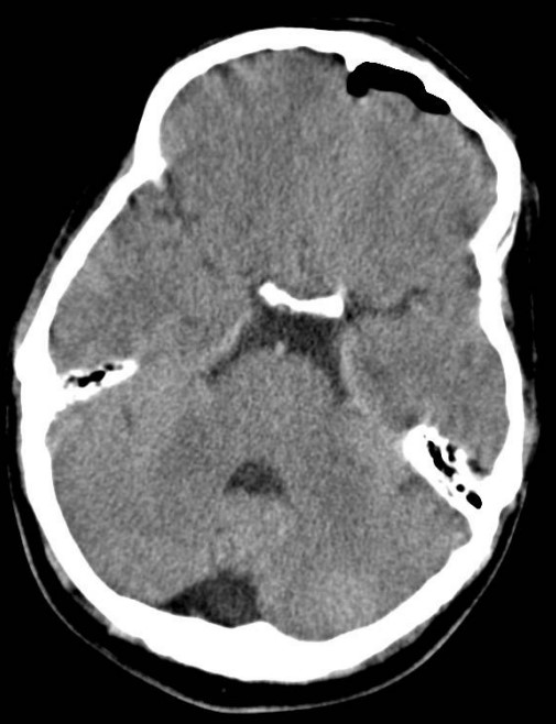
Figure 23: Unilateral pneumocephalus and enlarged occipital fossa, fourth ventricle, and suprasellar cistern (CT scan, axial section, brain window).
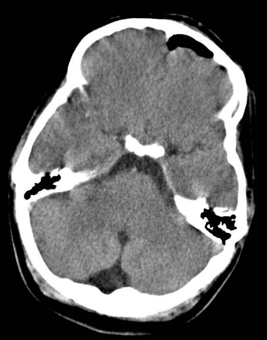
Figure 24: Unilateral pneumocephalus and enlarged occipital fossa, fourth ventricle, and suprasellar cistern (CT scan, axial section, brain window).
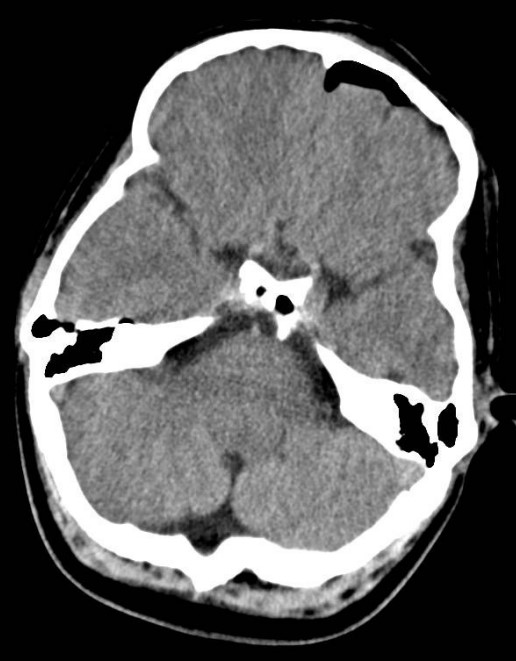
Figure 25: Unilateral pneumocephalus and enlarged occipital fossa, basilar cistern and circummesencephalic cistern (CT scan, axial section, brain window).
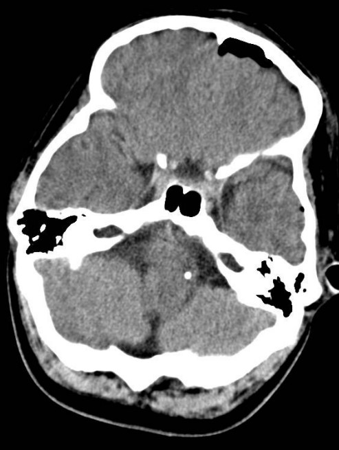
Figure 26: A calcified structure at pons level and the origin of the basilar artery.

Figure 27: Unilateral pneumocephalus anteriorly and vertebrobasilar dolichoectasia posteriorly (CT scan, axial section, brain window).

Figure 28: Unilateral pneumocephalus (CT scan, axial section, brain window).
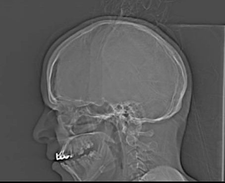
Figure 29: Pneumocephalus is seen on the topogram (CT scan, sagittal section for localization, topo gram).
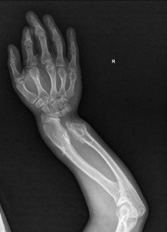
Figure 30: The upper limb affected by multiple bony exostoses (right side, x-ray, PA).
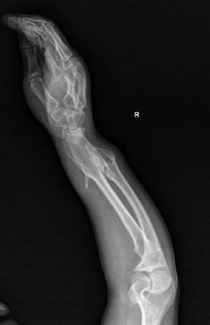
Figure 31: The upper limb affected by multiple bony exostoses (right side, x-ray, lateral).
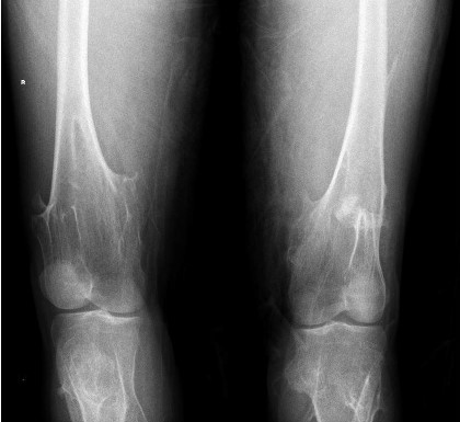
Figure 32: The lower limb affected by multiple bony exostoses (both knees, x-ray, PA).
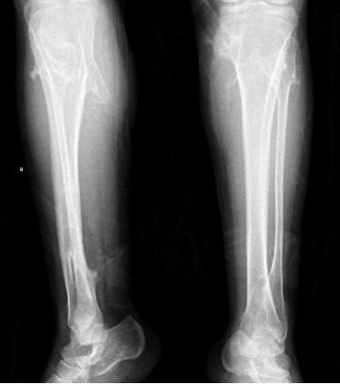
Figure 33: The lower limb affected by multiple bony exostoses (both legs, x-ray, AP).

Figure 34: The upper limb affected by multiple bony exostoses (both humerus, CT, axial section).
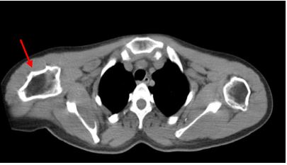
Figure 35: The upper limb affected by multiple bony exostoses (both humerus, CT, axial section).

Figure 36: The upper limb affected by multiple bony exostoses (both humerus and right scapula, CT, axial section).
Discussion
In tension pneumocephalus cases, a double-peaked silhouette of Mount Fuji is important neuroradiological sign [2]. The patient in this case did not lose conscious, suffer from neurological deficits, or have restlessness, but this case was stable. The mechanism of tension pneumocephalus is a ball-valve mechanism (one-direction valve to the subdural space) [2]. Pneumocephalus cases mostly are asymptomatic, but the patient had a severe headache. Air bubbles sign is seen in both pneumocephalus and tension pneumocephalus [3]. Pneumocephalus could progress to tension pneumocephalus. The rest of the findings, may or may not contributed to have a unilateral pneumocephalus. The patient has some hereditary conditions that could affect the healing process.
Conclusion
Mount Fuji sign is a classical sign that some cases do not have it. Unilateral pneumocephalus could be associated with hereditary conditions.
References
1. Chitrangada Singh. 2016. Post traumatic unilateral tension pneumocephalus [Internet]. Eurorad.
2. Ho ML, Eisenberg RL. 2014. Neuroradiology signs. McGraw Hill Professional. 67.
3. Sebastian B, Moideen J. 2018. Mount Fuji is Not as “Active” as We Think. Indian Journal of Neurosurgery. 7: 278-279.


