
-
Mail us:
editor@raftpubs.org
- ISSN : 2583-1534
Indexing & Abstracting
Full Text
Mini ReviewDOI Number : 10.36811/ojrmi.2021.110041Article Views : 2Article Downloads : 1
Importance of the oblique reconstruction of CT scan MPR formatting: A pictorial review
Abdulwahab Alahmari
Radiology Specialist, Radiology Department, Al-Namas General Hospital, Ministry of Health, Al-Namas City, Saudi Arabia
*Corresponding author: Abdulwahab Alahmari, Radiology Specialist, Radiology Department, Al-Namas General Hospital, Ministry of Health, Al-Namas City, Saudi Arabia, Tel: +966562428716; Email: afaa99@hotmail.co.uk
Article Information
Aritcle Type: Mini Review
Citation: Abdulwahab Alahmari. 2021. Importance of the oblique reconstruction of CT scan MPR formatting: A pictorial review. O J Radio Med Img. 4: 868-873.
Copyright: This is an open-access article distributed under the terms of the Creative Commons Attribution License, which permits unrestricted use, distribution, and reproduction in any medium, provided the original author and source are credited. Copyright © 2021; Abdulwahab Alahmari
Publication history:
Received date: 09 December, 2021Accepted date: 15 December, 2021
Published date: 20 December, 2021
Abstract
A common mistake among radiographers is reconstructing a CT scan and taking table top as a reference for the reconstruction. This paper, will mention in details what are some examples for oblique reconstruction with illustrated images.
Keywords: Oblique Reconstruction; Computed Tomography; Multi-Planer Reformat
Introduction
One of the common mistakes is reconstructing a CT scan and taking the table top as a reference of making the reconstruction even if the body is tilted to one side or the region of interest (ROI) is not normally placed in a straight way to be imaged and reconstructed directly. For example, the shoulder can’t be reconstructed from table top because it will make inaccurate cuts of the humerus which will not help in reading the CT scan. The landmark should be the glenoid cavity which lines should be parallel to in sagittal cuts and vertical on in axial cuts see (Figure. 1). The hip joint has the same rule apply to it. The oblique reconstruction is important to be used in the hip joint and the knee joint as seen in (Figure. 2&3). The azygous vein in the chest can visualize any pathology affect it by using an oblique reconstruction because it hides behind the right side of the heart and the great vessels see (Figure. 4). The heart is tilted to the left side in chest in most humans; therefore; all reconstructions should be made sagittal and coronal according the heart position itself see (Figure. 5). The temporal bone should be reconstructed according to the bone itself and can’t be reconstructed like a regular brain CT scan see (Figure. 6). The brain must be done based on the level of the base of the skull if not due to the patient condition or the need to do the scan as fast as possible, the reconstruction of the axial sections should be oblique and parallel to the base of the skull see (Figure. 7). The sagittal and coronal should be done vertical to the base of the skull. Reduction of angulation in brain CT will reduce the beam hardening artifact. If the patient duck his chin down, it will reduce the beam hardening artifact in foramen magnum. The oblique reconstruction will help in cutting the brain parallel to the base of the skull level. All these conclusions are as seen in (Table 1).
|
Table 1: The conclusions of published papers in this topic. |
|||
|
Author/s |
Year |
Sample size |
Conclusion |
|
Kimura et al [1]. |
2004 |
151 patients |
The oblique reconstruction can show hidden area in the chest like the mediastinum and costophrenic recesses. |
|
Massarotti et al [2]. |
2006 |
30 patients |
The oblique reconstruction provided additional information compared to the standard CT in the temporal lobe. |
|
Harris [3]. |
1981 |
1 animal (a dog) |
The oblique sections enhance the visibility of opacified coronary arteries |
|
Yin et al [4]. |
2015 |
55 patients |
The oblique reconstruction can help in taken an accurate measurement of the femoral neck torsion angle (FNTA). |
|
Oshina et al [5]. |
2018 |
18 patients |
The oblique reconstruction of the changed the intern-rater reliability from slight on sagittal, axial, coronal sections to fair for the oblique reconstruction in evaluating the spinal foramen stenosis. |
|
Ulla et al [6]. |
2009 |
20 patients |
The structures in the middle and inner ear are difficult to be evaluated on standard sections, the visualization can be improved on oblique reconstruction. |
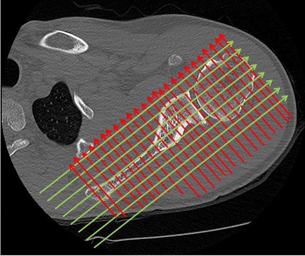
Figure 1: Reconstruction plans according to the shoulder joint, not to the table top.
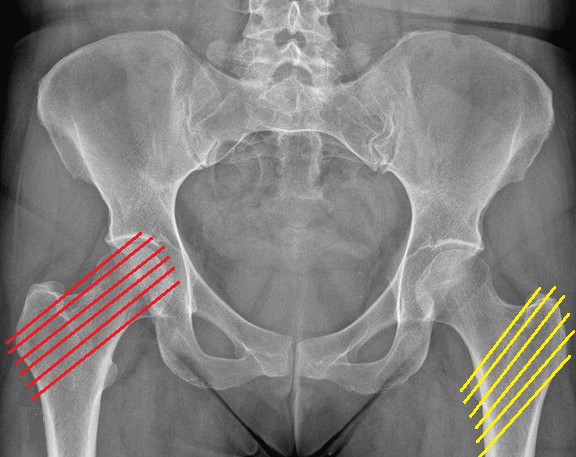
Figure 2: Oblique reconstruction plans should be along the femoral neck (red lines) and other oblique plans should be from the greater trochanter to the lesser trochanter (yellow lines).
Figure 3: The sagittal reconstruction should be made more tilted laterally in oblique fashion to cover some anatomical areas as seen in the planning on the axial section. The coronal should be vertical to these lines.
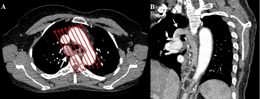
Figure 4: An oblique reconstruction of the great vessels which helped in showing azygous vein aneurysm (white arrow). The Figure. A. is an axial CT scan and Figure. B. is an oblique MPR CT scan.
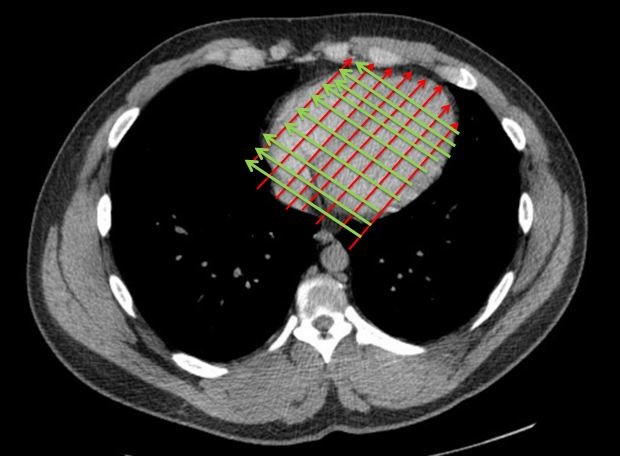
Figure 5: An oblique reconstruction of a CT scan MPR of the heart according to the heart position and not according to the whole chest.
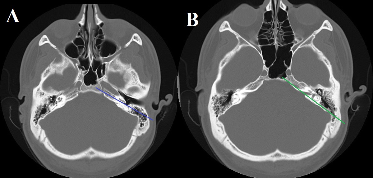
Figure 6: An oblique reconstruction should be parallel to the basal turn of the cochlea see (the blue line) on Figure. A. Other plans can be parallel to the temporal bone itself see (the green line) on Figure. B.
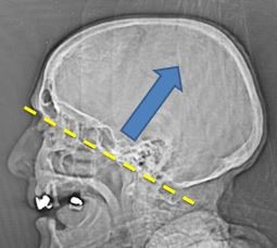
Figure 7: A CT tomogram showing how the brain should be reconstructed according to the base of the skull level. The cuts must be parallel to the yellow line from the base of the skull to the vertex following (the blue arrow). Notice the yellow arrow is below the base of the skull which indicate to take more below the base of the skull when a Radiographer is doing a brain CT.
Discussion
Some lung tumors and pathologies are required an oblique reconstruction because they are difficult locations in the chest like the mediastinum or costophrenic recess [1]. The oblique reconstructions can help in evaluating the coronary arteries [3]. Another paper indicates the usefulness of the oblique reconstruction in the temporal bone parallel to the basal turn of the cochlea [2]. The structure in the inner and middle ear were better visualized on oblique reconstruction [6]. In the spinal foramina, the oblique reconstruction allowed a better visualization which led to a better evaluation of the spinal foramina stenosis [5]. In the femur as well, the oblique reconstruction allowed a good visualization and accurate measurements of the femoral neck torsion angle [4].
Conclusion
The oblique reconstruction can give a better visualization of different part of the body which include; the temporal lobe, heart, mediastinum, large vessels in the chest, spine, hip, knee, etc. The oblique reconstruction will lead to a better evaluation, diagnoses, and obtaining an accurate measurement.
References
1. Kimura T, Naka N, Minato Y, et al. 2004. Oblique approach of computed tomography guided needle biopsy using multiplanar reconstruction image by multidetector-row CT in lung cancer. European journal of radiology. 2: 206-211. Ref.: https://pubmed.ncbi.nlm.nih.gov/15489081/ Doi: https://doi.org/10.1016/j.ejrad.2004.01.007
2. Mazziotti S, Arceri F, Vinci S, et al. 2006. Role of coronal oblique reconstruction as a complement to CT study of the temporal bone: normal anatomy. La radiologic medica. 2006 Jun; 111: 607-617. Ref.: https://pubmed.ncbi.nlm.nih.gov/16779546/ Doi.: https://doi.org/10.1007/s11547-006-0055-y
3. Harris LD. 1981. Identification of the optimal orientation of oblique sections through multiple parallel CT images. Journal of computer assisted tomography. 5: 881-887. Ref.: https://pubmed.ncbi.nlm.nih.gov/7320296/ Doi.: https://doi.org/10.1097/00004728-198112000-00018
4. Yin Y, Zhang L, Hou Z, et al. 2016. Measuring femoral neck torsion angle using femoral neck oblique axial computed tomography reconstruction. International orthopaedics. 40: 371-376. Ref,: https://pubmed.ncbi.nlm.nih.gov/26202020/ Doi: https://doi.org/10.1007/s00264-015-2922-4
5. Oshina M, Oshima Y, Tanaka S, et al. 2018. Utility of oblique sagittal reformatted and three-dimensional surface reconstruction computed tomography in foraminal stenosis decompression. Scientific reports. 8: 1-6. Ref.: https://pubmed.ncbi.nlm.nih.gov/30375504/ Doi: https://doi.org/10.1038/s41598-018-34458-9
6. Ulla MB, Vázquez F, Pumar JM, et al. 2009. Oblique multiplanar reformation in multislice temporal bone CT. Surgical and radiologic anatomy. 31: 475-479. Ref.: https://pubmed.ncbi.nlm.nih.gov/19190847/ Doi.: https://doi.org/10.1007/s00276-009-0463-5


