
-
Mail us:
editor@raftpubs.org
Indexing & Abstracting
Full Text
Case ReportDOI Number : 10.36811/ojor.2019.110004Article Views : 15Article Downloads : 22
Giant lipoma of the left neck: rare location
Anass Chaouki*, Ballage A, Choukry k, Mkhatri A, Abada R, Rouadi S, Roubal M and Mahtar M
Department of ENT, University Hospital Centre IBN ROCHD, North Africa
*Corresponding author: Anass Chaouki, Department of ENT, University Hassan 2 Casablanca, Moroocco, North Africa, Tel: +212618984627, Email: Anasschaouki.ac@gmail.com
Article Information
Aritcle Type: Case Report
Citation: Anass Chaouki, Ballage A, Choukry k, et al. 2019. Giant lipoma of the left neck: rare location. Open J Otolaryngol Rhinol. 1: 15-17.
Copyright:This is an open-access article distributed under the terms of the Creative Commons Attribution License, which permits unrestricted use, distribution, and reproduction in any medium, provided the original author and source are credited. Copyright © 2019; Anass Chaouki
Publication history:
Received date: 09 May, 2019Accepted date: 04 June, 2019
Published date: 06 June, 2019
Abstract
Giant lipoma is a rare cause of a large painless neck mass, these lipomas are usually found on the extremity, but rarely occur in the head and neck, computed tomography (CT) scan is very suitable for the diagnosis, Surgical excision is the best treatment. We describe a case of a man with a massive lipoma of the left neck, histological finding confirmed the diagnosis of spindle-cell lipoma, well managed with surgical excision.
Introduction
Lipomas are the most common type of soft tissue mesenchymal tumors, occur in the head and neck region in approximately 13% of cases, considered giant when it is weighs more than 1000 g or >10 cm in any dimension [1]. We report the case of a 60-year-old man with a massive lipoma of the left neck, with successful management.
Case report
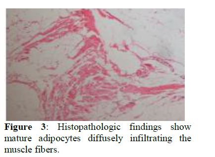
A 60-year-old man patient presented to the ENT department with 6 years’ history of increasing left sided neck mass, had been growing slowly, with an inability to fully turn his head, he denied any dysphagia, breathing difficulty, neurological symptoms or comorbidities. A large, smooth oval mass, 10 *7 cm in size (figure 1), was palpated along the left side of the neck. The skin of the mass showed normal, no erythema or inflammatory finding was detected. CT scan shows a low-attenuation massive mass involving the deep cervical fascia in the left side of the neck. There was no associated lymphadenopathy (figure 2). Under general anesthesia the patient underwent left neck dissection, a soft, friable and well encapsulated lipoma was discovered, extending into the prevertebral, jugular carotid and above clavicle plane and thus abutting the paravertebral muscles. In addition, the accessory nerve, brachial plexus, phrenic nerve, internal jugular vein, carotid artery and vascular element above the clavicle, at risk during the surgery, were identified and preserved. the mass was resected completely with the encapsulated sac of lipoma, muscles, nerve and vascular elements were respected, hemostasis ensured and no Plastic reconstruction was required. Histologic findings, fat tissues within muscle tissues benign spindle-cell with no malignant features confirmed the diagnosis (figure 3). Postoperatively, the patient demonstrated no neurological deficit and recovered the ability to fully turn his head and shoulder mobility. After 6 months’ follow-up the patient remind free of symptoms and no recurrence has been observed.
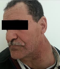
Figure 1: A large, smooth oval mass of the left neck.
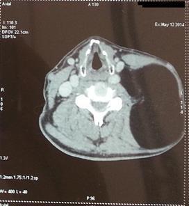
Figure 2: coronal CT scan showing a low-attenuation massive mass involving the deep cervical fascia.
Discussion
Lipomas are slow-growing, benign, soft tissue tumors that are typically asymptomatic, located subcutaneously and consist of mature fatty tissue; Lipoma in the head and neck area has been shown to be rare. It could occur in all age groups, preferentially in adults in their 30s to 60s [2]. Clinically, slowly growing asymptomatic mass or swelling with no palpable mass, it shows the characteristic of becoming soft and flat when surrounding muscles are relaxed. Paresthesia and nerve distribution neurological deficit due to nerve impingement can be encountered [3]. consistency can vary with the density of the fibrous tissue stroma specially when the engaged muscle is contracted. lateral neck masses are often seen in clinical practice, the first goal is to determine if the mass is malignant or benign; malignancies are more common in adult smokers older than 40 years; Other differential diagnoses for large lateral neck lumps include, lymphangioma, thyroid masses, lymphadenopathy and branchial cyst, but the key differential being liposarcoma and Metastatic squamous neck cancer especially with occult primary [4]. Metastatic squamous neck cancer with occult primary is a disease in which squamous cell cancer spreads to lymph nodes in the neck and it is not known where the cancer first formed in the body, Signs and symptoms include a lump or pain in the neck or throat, Tests that examine the tissues of the neck, respiratory tract, and upper part of the digestive tract are used to detect and diagnose metastatic squamous neck cancer and the primary tumor. For the diagnosis and differentiation of the tumor, ultrasonography, CT, and magnetic resonance imaging (MRI) may be used. Although ultrasonography shows hyper echogenicity, but it is influenced by the number of interfaces. computed tomography (CT) scan is very suitable for the confident identification of adipose tissues in lipomas. magnetic resonance imaging (MRI) is very useful for the diagnosis on T1-weighted images. [4,5] Management of lipoma will typically involve surgical excision; However, normal structures can be displaced, Care must therefore be taken to appreciate this anatomy and avoid these pitfalls [5]. The recurrence rate after treatment is 3% to 62.5%, and great differences were shown, depending on the investigators. This difference may be explained by incomplete resection [3,4].
References
- Copcu E, Sivrioglu N. 2005. Posterior cervical giant lipomas. Plast Recon Surg 115: 56-57.[Ref.]
- Singh M, Saxena A, Kumar L, et al. 2014. Giant lipoma of posterior cervical region. Case Rep Surg. 83.[Ref.]
- Virk JS. 2016. Massive lipoma of the posterior neck. BMJ Case Rep. 10: 1136.[Ref.]
- Won-Il Sohn. 2010. Intramuscular Lipoma of the Sternocleidomastoid Muscle. The Journal of Craniofacial Surgery. 21.[Ref.]
- Shane McTighe. 2014. Intramuscular lipoma: a review of the literature. Orthopedic Reviews. 6: 5618.[Ref.]


